The midsagittal section of the skull, a critical anatomical plane, unveils a comprehensive understanding of the skull’s intricate architecture. This section, meticulously dividing the skull into symmetrical halves, serves as a cornerstone for anatomical studies, clinical applications, and evolutionary comparisons.
Delving into the midsagittal section, we embark on a journey to explore the skull’s internal landscape, deciphering the intricate relationships between anatomical landmarks and their clinical significance. Comparative anatomy further enriches our understanding by highlighting evolutionary adaptations, while ongoing research and technological advancements continue to illuminate the potential of this section in medical research and practice.
Midsagittal Section of the Skull: Overview
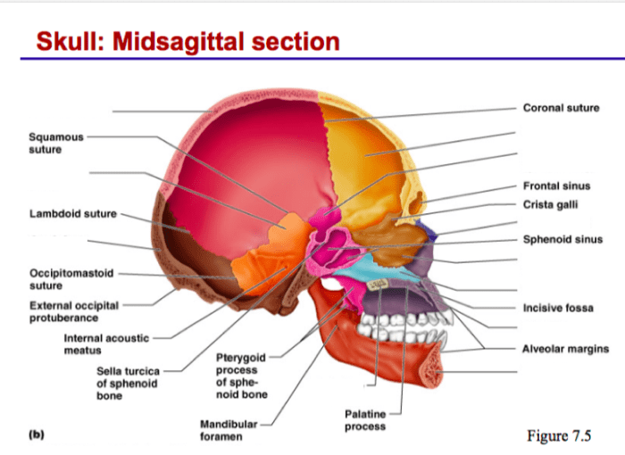
The midsagittal section of the skull is a vertical plane that divides the skull into left and right halves. It passes through the midline of the skull, from the nasion (the point where the nasal bones meet the frontal bone) to the opisthion (the point where the occipital bone meets the parietal bones).
The midsagittal section is an important anatomical landmark, as it provides a standardized reference point for describing the skull’s anatomy.
Midsagittal Plane
The midsagittal plane is a vertical plane that divides the body into left and right halves. It is defined by the median sagittal suture, which runs along the midline of the skull. The midsagittal plane is an important anatomical landmark, as it provides a standardized reference point for describing the body’s anatomy.
Importance of the Midsagittal Section
The midsagittal section of the skull is important for several reasons. First, it provides a standardized reference point for describing the skull’s anatomy. This is important for both clinical and research purposes, as it allows researchers to compare data from different individuals and studies.
Second, the midsagittal section can be used to assess the symmetry of the skull. This is important for diagnosing and treating conditions such as craniosynostosis, which is a condition in which the skull’s sutures close prematurely, resulting in an abnormal shape.
Third, the midsagittal section can be used to visualize the skull’s internal structures. This is important for diagnosing and treating conditions such as brain tumors and aneurysms.
Anatomical Landmarks
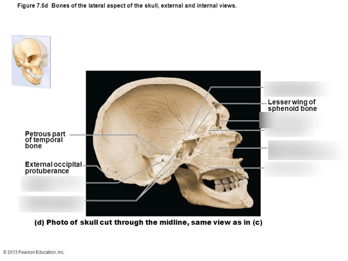
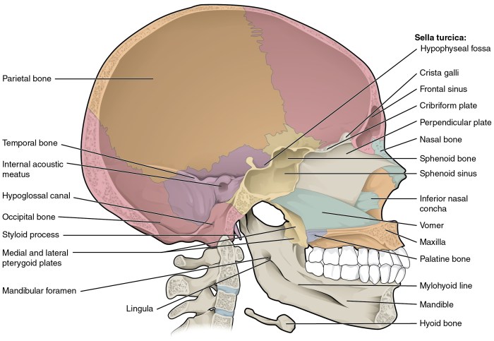
A midsagittal section of the skull, dividing it into left and right halves, reveals numerous significant anatomical landmarks. These landmarks provide valuable references for understanding the skull’s structure and its relationship with surrounding tissues.
The midsagittal section showcases a range of bony and soft tissue structures, including:
Cranial Bones
- Frontal bone: Forms the forehead and the roof of the orbits.
- Parietal bones: Paired bones that form the majority of the skull’s vault.
- Occipital bone: Forms the posterior aspect of the skull and the base of the cranium.
- Temporal bones: Located on either side of the skull, containing the organs of hearing and balance.
- Sphenoid bone: A complex bone located at the base of the skull, forming part of the orbital walls and supporting the pituitary gland.
- Ethmoid bone: A delicate bone located at the anterior base of the skull, forming part of the nasal cavity and supporting the olfactory bulbs.
Sutures
- Sagittal suture: A fibrous joint that connects the parietal bones along the midline of the skull.
- Coronal suture: A fibrous joint that connects the frontal bone to the parietal bones.
- Lambdoid suture: A fibrous joint that connects the parietal bones to the occipital bone.
Foramina and Canals
- Foramen magnum: A large opening at the base of the skull through which the spinal cord passes.
- Optic canal: A canal that transmits the optic nerve from the orbit to the cranial cavity.
- Internal acoustic meatus: A canal that transmits the facial and vestibulocochlear nerves to the inner ear.
Other Landmarks
- Falx cerebri: A fold of dura mater that separates the cerebral hemispheres.
- Tentorium cerebelli: A fold of dura mater that separates the cerebrum from the cerebellum.
- Pituitary fossa: A depression in the sphenoid bone that houses the pituitary gland.
- Nasal cavity: A space within the skull that houses the olfactory organs.
- Oral cavity: A space within the skull that houses the teeth and tongue.
Clinical Applications: Midsagittal Section: Midsagittal Section Of The Skull
The midsagittal section of the skull holds immense clinical significance, providing valuable insights into the anatomy and pathology of the head and brain. It serves as a crucial tool in medical imaging techniques, such as magnetic resonance imaging (MRI) and computed tomography (CT) scans.
These non-invasive imaging modalities allow clinicians to visualize the internal structures of the skull and brain, aiding in the diagnosis and treatment of various medical conditions.
Diagnostic Applications
The midsagittal section of the skull provides a comprehensive view of the brain’s midline structures, including the corpus callosum, thalamus, brainstem, and cerebellum. Abnormalities in these structures, such as tumors, cysts, or vascular malformations, can be detected through careful examination of the midsagittal section.
Additionally, the midsagittal section aids in evaluating the symmetry of the brain and detecting any midline shifts, which may indicate underlying pathology such as a mass lesion or hydrocephalus.
Treatment Planning, Midsagittal section of the skull
The midsagittal section of the skull plays a vital role in surgical planning and treatment of various neurological conditions. For instance, in cases of brain tumors, the midsagittal section helps determine the tumor’s size, location, and relationship to surrounding structures.
This information guides neurosurgeons in planning the surgical approach and minimizing the risk of damage to critical brain areas. Moreover, the midsagittal section assists in planning for procedures such as deep brain stimulation and stereotactic radiosurgery, where precise targeting of specific brain regions is essential.
Comparative Anatomy
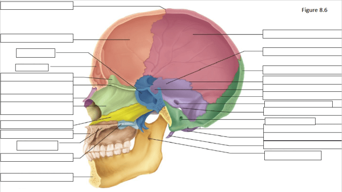
The midsagittal section of the human skull exhibits remarkable similarities and variations when compared to that of other vertebrates. Understanding these differences provides valuable insights into evolutionary adaptations and the functional diversity of the skull.
Humans vs. Non-human Primates
The midsagittal section of the human skull differs from that of non-human primates in several key aspects. Humans possess a larger and more globular braincase, accommodating the expanded cerebral cortex associated with advanced cognitive abilities. Additionally, the human skull features a more vertical forehead and a reduced supraorbital ridge, reflecting reduced reliance on the jaw muscles.
Humans vs. Carnivores
Compared to carnivores, the human midsagittal section exhibits a smaller facial region and a larger cranial vault. The reduced facial region reflects the omnivorous diet of humans, while the enlarged cranial vault accommodates the complex neural structures supporting higher-order cognitive functions.
Humans vs. Herbivores
Herbivores, such as ungulates, have a midsagittal section characterized by a large facial region and a relatively small braincase. This adaptation is associated with their grazing habits, requiring a robust jaw apparatus for processing plant material.
Evolutionary Adaptations
The variations observed in the midsagittal section of the skull among different vertebrates reflect their diverse evolutionary adaptations. The enlarged braincase and reduced facial region in humans are adaptations for advanced cognition and tool use. The smaller facial region and larger cranial vault in carnivores support their predatory lifestyle, while the large facial region and small braincase in herbivores facilitate their herbivorous diet.
Research and Advancements: Midsagittal Section
Recent research in the field of medical imaging has focused on developing advanced techniques to enhance the visualization and analysis of the midsagittal section of the skull. These advancements have led to a better understanding of the anatomy and pathology of this region, aiding in the diagnosis and treatment of various medical conditions.
Imaging Advancements
One significant advancement has been the development of high-resolution computed tomography (CT) and magnetic resonance imaging (MRI) techniques. These technologies provide detailed cross-sectional images of the skull, allowing for precise visualization of the midsagittal section. Additionally, three-dimensional (3D) reconstruction techniques have been developed, enabling the creation of virtual models of the skull for further analysis and surgical planning.
Clinical Applications
The improved visualization of the midsagittal section has led to several clinical applications. In neurosurgery, it assists in the planning and execution of complex procedures such as tumor removal, aneurysm clipping, and skull base surgery. In orthodontics, the midsagittal section provides valuable information for assessing jaw alignment and planning corrective treatments.
Moreover, it is useful in forensic anthropology for facial reconstruction and identification.
Future Directions
Future research and advancements in the field of midsagittal section imaging are expected to focus on further refining existing techniques and developing novel approaches. These may include the integration of artificial intelligence (AI) and machine learning algorithms for automated analysis and interpretation of imaging data.
Additionally, there is potential for the development of personalized imaging protocols tailored to individual patient anatomy and pathology, leading to more accurate and effective diagnosis and treatment.
FAQ Overview
What is the midsagittal plane?
The midsagittal plane is a vertical plane that divides the body into symmetrical left and right halves.
What are the major anatomical landmarks visible in a midsagittal section of the skull?
The major anatomical landmarks visible in a midsagittal section of the skull include the frontal bone, parietal bone, occipital bone, sphenoid bone, ethmoid bone, nasal bone, maxilla, mandible, and teeth.
What is the clinical significance of the midsagittal section of the skull?
The midsagittal section of the skull is clinically significant because it provides a clear view of the skull’s internal structures, which can be useful for diagnosing and treating various medical conditions, such as skull fractures, brain tumors, and sinus infections.
